OEM Collimators,
manufactured to drawing.
Custom collimator assemblies for SPECT and gamma-camera systems — built to the customer's drawing, BOM, and inspection criteria. Programs span new platform development, current-generation supply, and ongoing service replacement for legacy installations.
OEM collimator
manufacturing.
Complete assemblies for SPECT and gamma-camera platforms. Channel geometry, septa configuration, hole length, format size, and mounting interface — all produced to the customer drawing. Across new platform development, current-generation supply, and ongoing service replacement.
- Geometries
- Parallel-hole, fan-beam, pinhole, rectangular-hole.
- Energy classes
- LEHR, LEHRS, LEUHR, MEGP, HEGP.
- Programs
- OEM, custom, replacement, service supply.
- Quality
- ISO 9001:2015 certified by DEKRA. Full lot traceability.
Why the collimator
decides the image.
A gamma camera cannot focus its photons. At the energies of SPECT and planar nuclear medicine imaging there is no lens and no mirror — the only way to form an image is to absorb every photon travelling on the wrong path. That is the task of the lead collimator: a matrix of precision channels admitting only the rays that arrive along the designed direction. It is the single component that defines what the detector is able to see.
Every collimator is a negotiated trade-off. Narrower, longer channels sharpen spatial resolution but admit fewer photons; wider, shorter ones collect more counts at the cost of detail. Septal thickness follows the isotope — the 364 keV photons of ¹³¹I demand far more lead between channels than the 140 keV of ⁹⁹ᵐTc. Matching channel geometry, septa, and hole length to the clinical task is the core discipline of SPECT collimator design.
The physics is unforgiving of manufacturing error. A clinical acquisition uses only a small fraction of the photons a patient emits, so small geometric deviations — channel angulation, septal thickness, pitch — translate directly into visible artifacts and lost resolution. This is why collimator manufacturing is measured in hundredths of a millimetre, and why every assembly is inspected against its drawing before it reaches a detector head.
Geometry
portfolio.
Four standard geometries, each engineered for a different clinical question. Channel geometry, septa, and interface are manufactured to the customer drawing.
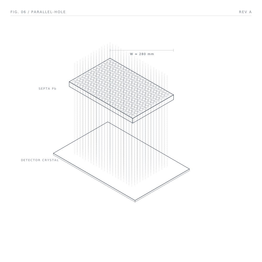
Identical channels perpendicular to the detector. Only true-normal photons survive the septa; the image is geometrically faithful at 1:1.
Whole-body bone, lung perfusion, renal, brain SPECT.
Parallel-Hole
The general-purpose SPECT geometry. Low, medium, and high energy across the diagnostic isotope range.
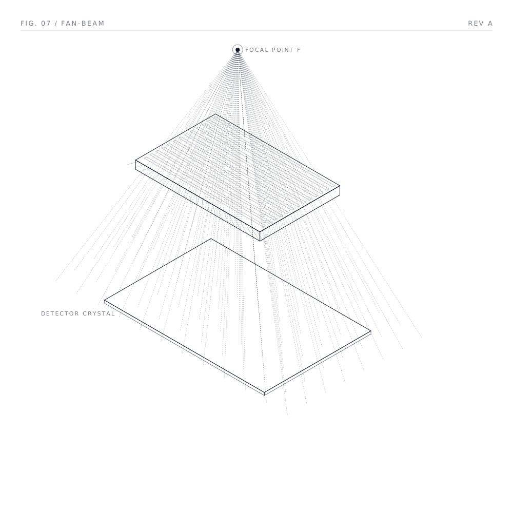
Channels converge toward a focal line; the projection magnifies in one axis while preserving 1:1 in the other — trading field of view for detail.
Brain SPECT (DaTscan, perfusion), small-organ studies.
Fan-Beam
Converging-geometry optics for brain SPECT and small-organ acquisitions. To customer focal length.

Photons cross at a single aperture and form an inverted image on the detector. Magnification is geometric — set by focal and object distance.
Thyroid, parathyroid, sentinel node, paediatric SPECT.
Pinhole
Magnifying single-aperture optics for the highest spatial resolution on small targets.
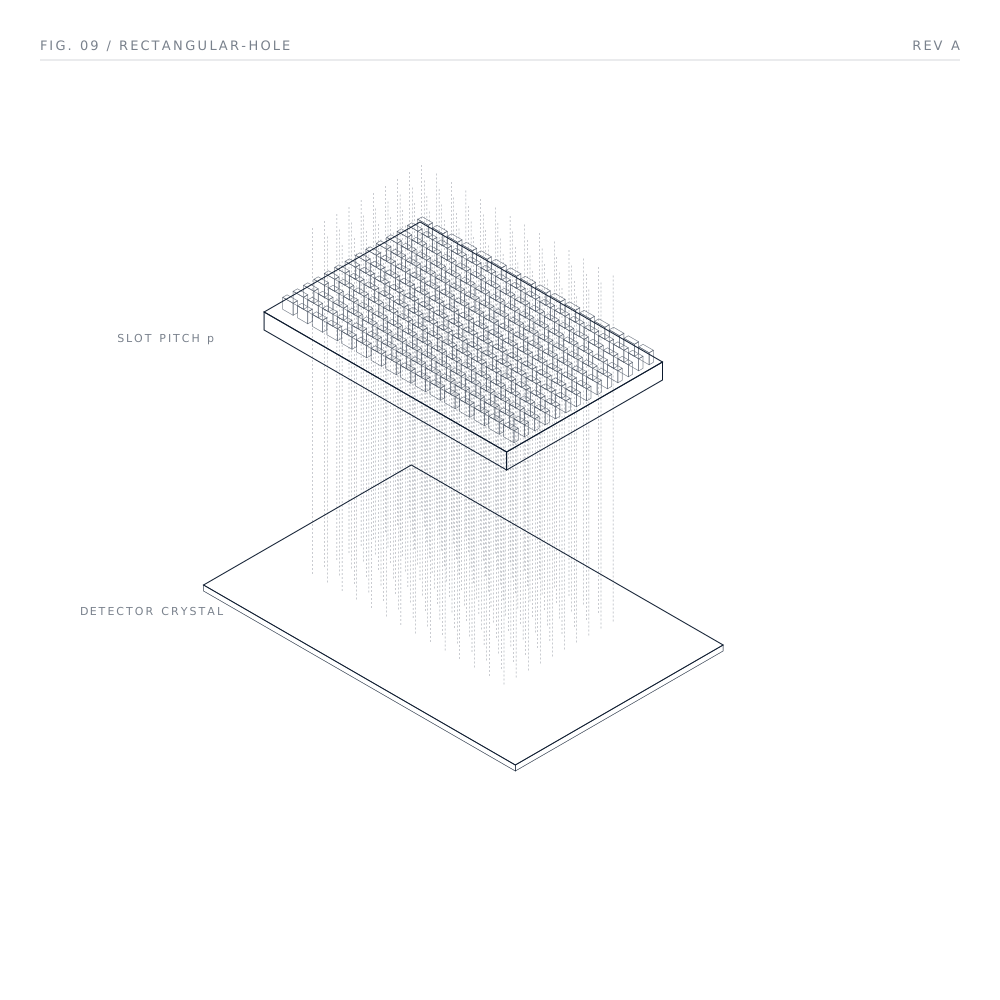
Rectangular-section channels perpendicular to the detector. The long axis raises geometric efficiency for slot-shaped targets; the short axis preserves resolution.
Whole-body planar, lymphoscintigraphy, high-throughput.
Rectangular-Hole
Slot-channel geometries built to OEM detector pitch. High-sensitivity acquisitions.
Energy
classes.
Three principal classes, each defined by the gamma-ray energy it must stop. Customer terminology varies (LEHR, LEHRS, LEUHR, MEGP, MELP, HEGP, HEHR): underneath, three energy bands cover the full diagnostic and therapeutic isotope range.
| Class | Energy | Isotopes | Clinical use |
|---|---|---|---|
| LELow energy · LEHR / LEHRS / LEUHR / LEGP | 30 – 200 keV | ⁹⁹ᵐTc · ²⁰¹Tl ¹²³I · ¹¹¹In | General-purpose SPECT — bone, lung perfusion, brain, renal, cardiac. |
| MEMedium energy · MEGP / MELP | 200 – 400 keV | ⁶⁷Ga · ¹¹¹In ¹⁷⁷Lu · ¹²³I high-energy | Therapy monitoring, sentinel-node localisation, and infection imaging. |
| HEHigh energy · HEGP / HEHR | 350 – 600 keV | ¹³¹I · ¹⁸F theranostic isotopes | Post-therapy imaging, ¹³¹I scans, and emerging theranostic protocols. |
Manufactured for the SPECT and gamma-camera platforms
in active clinical use today.
SPECT collimators Discuss a
collimator program.
Send drawings, BOM, or a target specification. We will review feasibility, materials, and inspection criteria. Response within two business days.